
Advanced Light Microscopy
Contacts
Staff

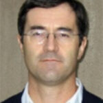
Christopher O'Connell, Ph.D.
Facility Director
- Biology Physics Building, Rm G05C
- coconnell@uconn.edu
- 860.486.3271
Faculty Scientific Advisers

Juliet Lee, Ph.D.
Associate Professor
- Biology/Physics Building 306
- juliet.lee@uconn.edu
- 860.486.4332

Akiko Nishiyama, Ph.D.
Professor
- Pharmacy/Biology Building 631
- akiko.nishiyama@uconn.edu
- 860.486.4561
Location
Campus Address
Mailing Address
Advanced Light Microscopy Facility
Center for Open Research Resources and Equipment
75 North Eagleville Road
Storrs, CT 06269-1149
Instrumentation
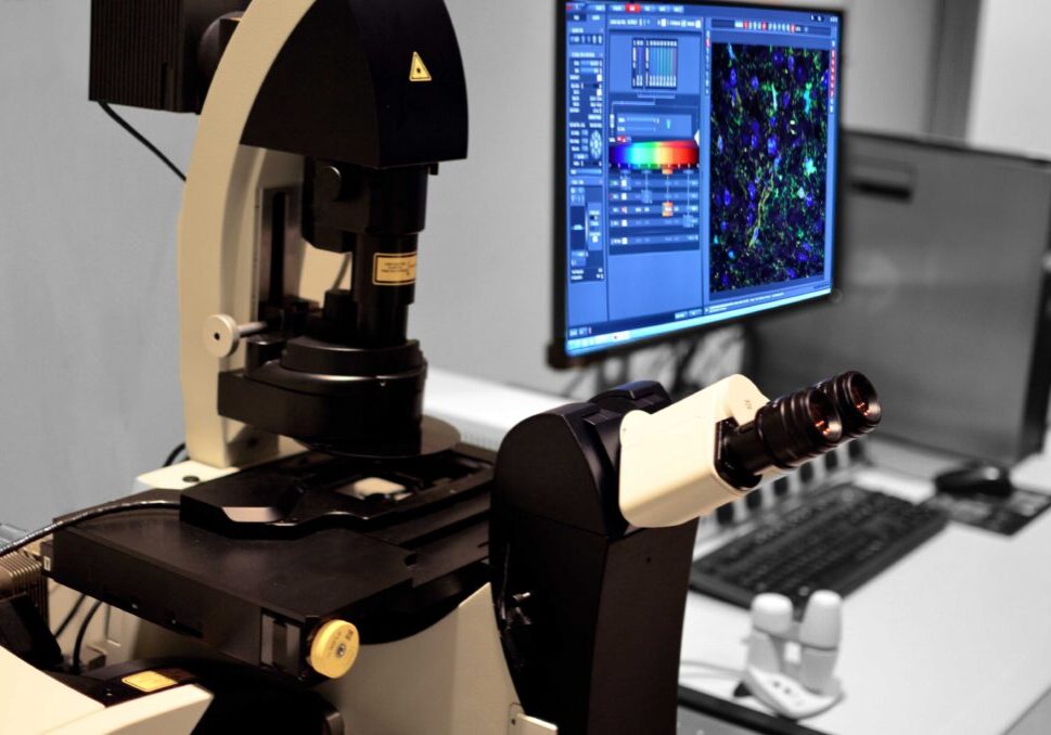
Leica SP8 Spectral Confocal
This is an inverted confocal microscope with five filter-free spectral and individually regulatable channels including four standard PMT detectors and one high-sensitivity HyD detector with photon counting capacity. The scanner also includes both standard galvo scan mirrors and an optional 8 KHz resonant scanner for high speed imaging. Use of an acusto-optical beamsplitter (AOBS) instead of traditional dichroic mirrors makes this microscope also suitable for reflected light microscopy applications. The instrument is housed in the Bioscience Electron Microscopy Laboratory located in the Biology and Physics Building (BPB), room G10.
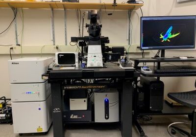
Nikon AXR Confocal + TIRF
The Nikon AXR is a point scanning confocal built around the inverted Ti2E microscope. The large 25 mm field of view and resonant scanner are optimized for rapid imaging of samples. The DUX-VB confocal detector features 4 sensitive GaAsP detectors that can be flexibly configured for a range of dyes via spectral detection. 6 laser lines provides a range of excitation options for a variety of fluorescent dyes. In addition to confocal microscopy, the system has an iLAS2 illumination module for TIRF imaging. This device can perform simultaneous TIRF imaging and photostimulation. TIRF, widefield, and transmitted light images are collected on a Photometrics Prime 95B sCMOS camera. The instrument is located in the Biology and Physics Building (BPB), room G05D.
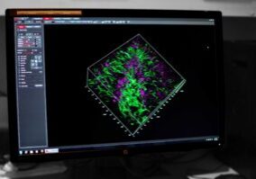
Image Analysis Workstation
The Image Analysis Workstation is a powerful computer dedicated for image analysis and visualization. Several software packages are installed to support a variety of data formats.
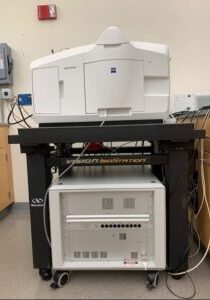
Zeiss Lightsheet 7
The Zeiss Lightsheet 7 microscope is designed for fast, gentle 3D/4D imaging of samples labeled with fluorescent probes. Both live and fixed samples can be imaged. Samples are lowered from above into an imaging chamber containing media of the appropriate refractive index. It is located in BPB G05.
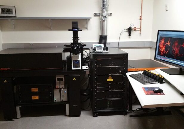
Abberior Instruments 3D STED
This is an inverted confocal microscope that uses stimulated emission depletion (STED) of fluorescent dyes to obtain super resolved images. There are four pulsed excitation lasers and two pulsed STED depletion lasers for imaging. The instrument is capable of 2D or 3D super resolution imaging using spatial light modulators to shape the depletion beams in the x-y and z axes. The instrument is housed within the Bioscience Electron Microscopy Laboratory located in the Biology and Physics Building (BPB), room G11.
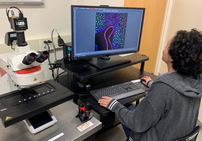
Leica Thunder Imager
The Leica Thunder Imager is an upright microscope for fluorescence, transmitted light, and true color imaging . With a stage capable of holding up to 8 slides, this instrument is optimized for high throughput imaging of cells and tissue sections. Fluorescent images can be processed with Leica’s computational clearing and deconvolution to remove out of focus blur, enhance contrast, and sharpen details. The software Navigator function can be used for rapidly generating sample overviews for subsequent selection of areas of interest. The instrument is located in the Biology and Physics Building (BPB), room G05D.
Services and Rates
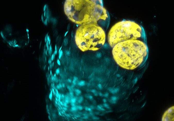
Overview
The Advanced Light Microscopy Facility provides training and access to advanced light microscopy systems at an hourly rate. In addition, we are available to consult with and support users at every stage of a project including: experimental design, sample preparation, image acquisition, analysis, and data preparation.
- Consultation: We are available to meet with current and potential users to advise on sample preparation and the selection of the instrument that best serves the research requirements.
- Training: All users are provided with training that covers facility policies and details of instrument operation.
- Instrument Access: Researchers are able to independently use the instruments after training, or they can work with the Director in assisted sessions. See Instrumentation for current rates.
- Support: Our expertise is always available to help maximize the impact of the instruments on users’ studies through further consultations or by scheduling assisted sessions with the Director on the microscope.
- Education: The Facility gives workshops and lectures on microscopy applications and image analysis to advance the microscopy knowledge of the research community.
- Billing: Monthly usage is tabulated and billing is executed by the Center for Open Research Resources and Equipment (COR2E).