Reverse Engineering Fabrication Inspection and Non-Destructive Evaluation (REFINE)
Contacts
Staff
Location
Campus Address
Innovation Partnership Building
Mailing Address
159 Discovery Drive
Storrs, CT 06269
Instrumentation

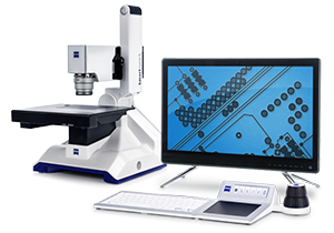
ZEISS Smartzoom 5
Microscope for QC and Failure Analyses
Smartzoom 5 is your smart digital microscope - ideal for QC/QA and Failure Analysis applications in virtually every field of industry. Fully automated and equipped with dedicated workflows, it’s so simple to operate, even untrained users will produce excellent results. With integrated overview camera for Google Earth like navigation makes the area of interest fast and easy to locate. A wide magnification range of 10x-2000x with 10x Zoom covers a variety of applications for 2D and 3D imaging and measurement.

ZEISS Smartproof 5
Your Integrated Confocal Microscope for Surface Imaging
The versatile ZEISS Smartproof 5 widefield confocal microscope is your integrated non-contact system for surface analysis: fast, precise and repeatable. Put it to work on a wide range of industrial applications - such as surface roughness and topographic measurement with nanometer resolution that comes up daily in QA/QC, failure analysis and R&D labs.

ZEISS ORION Nanofab
ZEISS ORION NanoFab is a unique focused ion beam instrument capable of generating helium, neon, and gallium ion beams. Based on the Gas Field Ion Source (GFIS) technology, the instrument creates an extremely collimated, highly coherent ion beam that can be used for highest resolution imaging with probe sizes of less than 0.5nm and for precise nanofabrication down to a dimension of about 5nm. This allows for the fabrication of structures such as plasmonic devices and nanopores, patterning of 2D materials, or ion beam lithography with feature sizes well below of what can be achieved with conventional gallium FIBs. The use of inert ion species also eliminates metallic contamination during the milling process and enables deposition of ultra-fine high quality metal or insulator lines. The addition of a gallium focused ion beam column allows for large volume material removal at high speeds.

ZEISS Crossbeam
An FE-SEM/FIB/fiber laser system capable of high-resolution 3D analytics. Combines multi-scale milling and imaging.

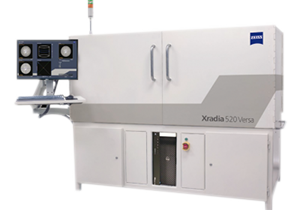
ZEISS Xradia 520 Versa
Unlock new degrees of versatility for your scientific discovery and industrial research with the ZEISS Xradia 520 Versa 3D X-ray microscope; the most advanced model in the Xradia Versa family. Building on industry-best resolution and contrast, Xradia 520 Versa expands the boundaries of non-destructive imaging for breakthrough flexibility and discernment critical to your research. Innovative contrast and acquisition techniques free you to seek – and find – what you have never seen before. Move beyond exploration and achieve discovery. Innovative contrast and acquisition techniques free you to seek – and find – what you have never seen before.