
Nuclear Magnetic Resonance Spectroscopy
Nicholas Eddy, Ph.D.
- nicholas.eddy@uconn.edu
- 860.486.2568
Capri Price, Ph.D.
- capri.price@uconn.edu
- 860.486.5851
Location
Campus Address
Mailing Address
25 King Hill Road, Unit 3136
Storrs, CT 06269-3136
Instrumentation
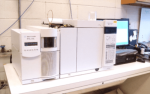
Gas Chromatography Mass Spectroscopy – Agilent GCMS Systems
A sample is injected into a heated inlet, then the component molecules of the sample mixture are separated by interaction with a coated capillary column in the gas chromatograph. The separated molecules are detected with a mass spectrometer, allowing for identification. Contact: Capri Price
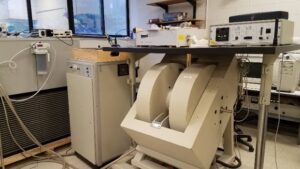
Electron Paramagnetic Resonance – Bruker EMX EPR
Samples are placed into a microwave cavity while the external magnetic field is changed. Electrons will undergo a change in alignment at the resonant frequency in the microwave field. This technique can be done in a quantitative method. Contact: Nicholas Eddy
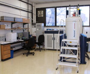
Nuclear Magnetic Resonance - Bruker Avance III 400 WB
The Bruker Avance III 400 MHz Wide Bore (WB) NMR spectrometer is a three-channel system dedicated for solid samples with Magic Angle Spinning (MAS) and Pulse Field Gradient capabilities. The spectrometer is equipped with a triple resonance HXY 4 mm MAS probe and a triple resonance HFX 2.5 mm MAS probe for solids with VT from – 50 to +150 degrees Celsius running experiments with Nitrogen gas. This instrument also has a double resonance H(F)X 5 mm Doty Scientific Pulse Field Gradient (PFG) probe with a Z axis Great 1/40 gradient amplifier for diffusion studies of liquid and solid samples. Contact: Nicholas Eddy
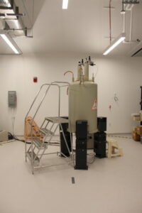
Nuclear Magnetic Resonance – Bruker DMX 500
Solution state NMR is used for determining the average structure of one molecule. It provides information on chemical structure and quantifiable amounts present in solution. This type of NMR can detect ppm levels of nuclei in solution. Contact: Nicholas Eddy
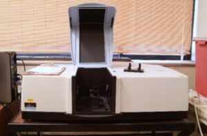
UV‐VIS‐NIR Spectroscopy – Perkin Elmer Lambda 1050
Exposure to desired discrete wavelength(s) tests the sample’s ability to absorb/transmit light. This technique can be used to determine the band gap of a material. Contact: Capri Price
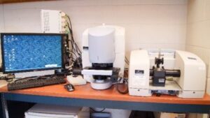
Molecular Analysis – Perkin Elmer Spotlight 400 Imaging System (micro-FTIR)
A sample is illuminated with infrared light, the absorption of which causes molecular vibrations in the sample. These vibrations are characteristic of functional groups in the sample. A representative spectrum is shown to the left with some of the evident functional groups denoted. FTIR is not able to determine connectivity of functional groups. Contact: Capri Price
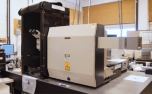
Raman Spectroscopy – Renishaw Ramascope 2000
Stimulating the sample with a laser causes a phenomenon called Raman scattering. This inelastic scattering allows for identification of functional groups and is considered complementary to infrared spectroscopy. In some cases it can also provide information on crystallinity, purity, stress & strain or defects. Contact: Capri Price
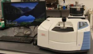
Molecular Analysis – Thermo Fisher Nicolet iS20
A sample is illuminated with infrared light, the absorption of which causes molecular vibrations in the sample. These vibrations are characteristic of functional groups in the sample. A representative spectrum is shown to the left with some of the evident functional groups denoted. FTIR is not able to determine connectivity of functional groups. Contact: Capri Price
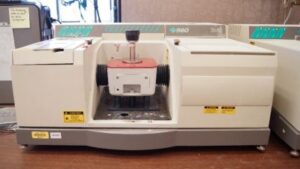
Molecular Analysis – Thermo Fisher Nicolet Magna 560
FTIR-ATR is capable of rapidly determining the major organic chemical species present in a sample, with a detection limit of 1-5% by weight. FTIR-GA is used for analyzing molecular layers on a reflective surface. FTIR-Transmission can provide a greater signal-to-noise ratio in some cases than attenuated total reflectance. Contact: Capri Price