Flow Cytometry
Contacts
General Contact
860.679.3463
Instrumentation

BD FACSymphony A5 SE
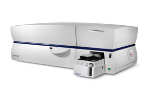
There are two identical BD FACSymphony A5 SE cell analyzers which have five lasers and 48 algorithmically optimized fluorescence detectors for cellular analysis. This instrument can be used for both conventional and spectral flow cytometry because it uses additional high-sensitivity detectors and new software functionality to detect the full spectrum of emitted light and unmix spectrally live in BD FACS Diva software, all while retaining the flexibility of familiar compensation-based workflows. Both instruments have an available 96-or 384-well plate reader attached.

BD FACSymphony S6 Cell Sorter
The FACSymphony S6 SE has five lasers and 48 algorithmically optimized fluorescence detectors for cellular analysis. This instrument can be used for both conventional and spectral flow cytometry because it uses additional high-sensitivity detectors and new software functionality to detect the full spectrum of emitted light and unmix spectrally live in BD FACS Diva software, all while retaining the flexibility of familiar compensation-based workflows. The sorter is capable of separating 6 distinct cellular populations and has the capacity to sort single cells into 96- or 384-well plates or onto glass slides. The FACSymphony S6 SE cell sorter is installed in a biosafety cabinet and is equipped with an aerosol containment unit to allow sorting of biohazardous materials.

Becton-Dickinson LSR II
The LSR II-B cell analyzer has five lasers and is able to detect 20 parameters (18 fluorescence plus forward and side scatter). LSR II-B is considered a special order research products (SORP) and was customized for ongoing research at UConn Health.

Becton-Dickinson FacsAria II
The FACS Aria II high-speed cell sorter has five lasers. and can detect up to 18 parameters (16 fluorescence colors plus forward and side scatter). The Aria II cell sorter is installed in a biosafety cabinet and is equipped with an aerosol containment unit to allow sorting of biohazardous materials. Additionally, the Aria II cell sorter is equipped with an automated cell deposition unit (ACDU) that allows for sorting into plates or glass slides. The Aria II sorters is considered a special order research products (SORP) and was customized for ongoing research at UConn Health.

BioRad ZE5
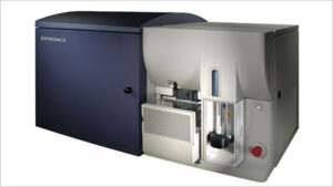
The ZE5 Cell Analyzer has 5 lasers and allows for 27 fluorescence parameter flow cytometry analysis. It integrates a universal plate loader that accepts single tubes, tube racks, or 96- and 384-well plates. It has integrated sample vortexing, sample temperature control, and volumetric cell counting. Forward scatter can be detected from both the 488nm and 405nm lasers which may allow for better detection of small particles.

Accuri C6
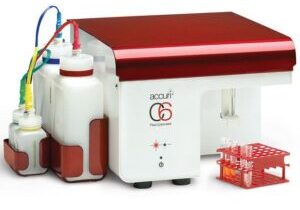
Located in room E3004, this simple high throughput system is equipped with a blue and red laser, two light scatter detectors, and four fluorescence detectors with optical filters optimized for the detection of FITC, PE, PerCP, and APC. A compact optical design, fixed alignment, and preoptimized detector settings make the system easier to use. Data is digitally collected over a wide dynamic range of 7.2 decades (16 million channels of digital data), making all data available to users as needed. Gating strategies and fluorescence compensation values can be set before, during, or after data collection.

Amnis ImagestreamX Mark II
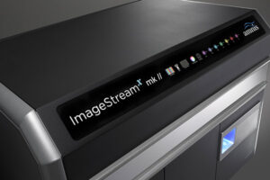
The revolutionary ImageStream®X Mark II Imaging Flow Cytometer combines the speed, sensitivity, and phenotyping abilities of flow cytometry with the detailed imagery and functional insights of microscopy. This unique combination enables a broad range of applications that would be impossible using either technique alone.
This instrument produces multiple high-resolution images of every cell directly in flow, including brightfield and darkfield (SSC), and up to 10 fluorescent markers with sensitivity exceeding conventional flow cytometers. Applications include the study of cell-cell interactions, phagocytosis, apoptosis and autophagy, the characterization of circulating tumor cells, and many others.
Services
Comprehensive List of Equipment in the Flow Cytometry Facility at UConn Health
BD FACS Symphony A5 Spectral flow cytometry cell analyzer (x2)
BD FACS Symphony S6 Spectral flow cytometry cell sorter
BD LSRII flow cytometry cell analyzer
BD FACS ARIA flow cytometry cell sorter
BD Accuri C6 flow cytometry cell analyzer
Amnis Imagestream X Mark II imaging flow cytometer
Bio-Rad ZE5 flow cytometry cell analyzer
Sorvall tube and plate centrifuge
Eppendorf 1.5mL tube centrifuge
FlowJo flow cytometry analysis software workstations
BD FACS DIVA flow cytometry analysis software workstation
Miltenyi Auto-MACS magnetic cell separator
Miltenyi Octo MACS magnetic cell separation magnet
Miltenyi Midi MACS magnetic cell separation magnets (x3)
Miltenyi Vario MACS magnetic cell separation magnet
Campus Address
E Building, Room E6014
UConn Health Center
Mailing Address
263 Farmington Ave
Farmington, CT 06030