MicroCT Imaging Facility
Contacts
Instrumentation

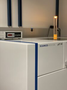
Scanco µCT50 (Excised Specimens)
- Max spatial resolution: 2 µm (10% MTF); Max nominal resolution (voxel size) 0.5 µm
- Max voltage: 100 kVp; Max current: 200 µA at all energies; 3400 x 1200-element detector
- Optional compression/tension stage can apply up to 500N load (±1%) and 9mm displacement (±0.02mm)
- Optional temperature stage allows small samples to be heated/cooled ±15°C from ambient temperature
- Appropriate for all specimens compatible with µCT40
For up to date rates please visit here

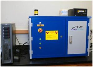
Scanco µCT40 (Excised Specimens)
- Max spatial resolution: 8 µm (10% MTF); Max nominal resolution (voxel size) 3 µm
- Max Voltage: 70 kVp; Max current 177 µA at lowest energy; 2048 x 256-element detector
- Field of view: 10-40 mm diameter × 70 mm length
- Quantitative analysis of bone architecture (cortical and trabecular measurements)
- Rodent bone morphometry, fracture repair, tissue engineering constructs
- Contrast-enhanced imaging of tumors and vasculature
- High-contrast imaging of heavy metal-stained organs, embryos, marrow adipocytes
- Select applications in materials science, pharmaceutical manufacturing, and museum/educational pieces
- Scan time scales with resolution and field of view height. As an example, a set of 4 full-length mouse femurs, at 16 µm resolution, takes approximately 110 minutes of scan time. The same scan at 8 µm resolution takes twice as long.
For up to date rates please visit here

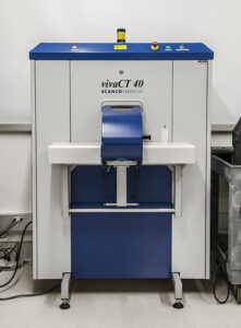
Scanco VivaCT40 (Live Animals)
- Field of view: 20-40 mm diameter × 140 mm length
- Spatial resolution: 10 µm to 40 µm
- Longitudinal quantitation of bone morphometry (best suited for cranial imaging)
- Contrast-enhanced imaging of vasculature, tumors, etc.
- Primarily used for in vivo imaging of mice (or small rats) under isoflurane anesthesia.
- IACUC approval is required for all procedures involving live animals.
For up to date rates please visit here
TA Instruments Electroforce 3200 (Biomechanical Testing)
- Currently set up for 3-point bending (failure testing) of mouse long bones.
- High-resolution, low force, small displacement
- Axial and rotary actuators (tension, compression, flexure, torsion)
- Resolution and control to 10 µm in position, 50 mg force
Bose SLM090 (Biomechanical Testing)
- Currently set up for torsional testing of mouse long bones (fracture healing).
- High torque servo motor: up to 56 lbf-in and up to 4000 rpm
- 2 Nm MRT flange-style reaction torque transducer
CSM Nanoscratch (Biomechanical Testing)
- Please inquire if you are interested in using this instrument.
Other Project Support Infrastructure
- Sherline 4400 3" x 17" tabletop CNC lathe: not set up--contact if interested
- Sherline 2000 8-direction tabletop CNC mill: not set up--contact if interested
- Various rotary tools (Dremel)
Mailing Address
Dept. of Orthopaedic Surgery, MC 4037
263 Farmington Avenue
Farmington, CT 06030-4037