Core Incentive Plan (CIP) Program
UConn Health HCRAC Facilities
To apply, please reach out to the appropriate core director below:
UConn Health HCRAC Facilities
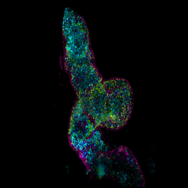
The CCAM Microscopy Facility provides the UConn Health research community as well as other academic and industrial institutes, access to its state-of-the-art equipment for quantitative fluorescence imaging applications. Within the facility, there are two laser scanning confocal microscopes equipped with 34-channel high-efficiency QUASAR detectors. One of these microscopes supports nonlinear optical (also known as 2-photon) excitation, fluorescence correlation spectroscopy (FCS), and fluorescence lifetime imaging microscopy (FLIM). Additionally, there is a light sheet microscope available for micro/meso scale 3D imaging. Furthermore, the facility recently acquired a state-of-the-art Zeiss Elyra 7 microscope that is capable of total internal reflection fluorescence microscopy (TIRFM) and supports several super-resolution techniques such as lattice structured illumination microscopy (SIM), stochastic optical reconstruction microscopy (STORM), 3D photoactivated localization microscopy (PALM), and DNA points accumulation for imaging in nanoscale topography (DNA-PAINT).
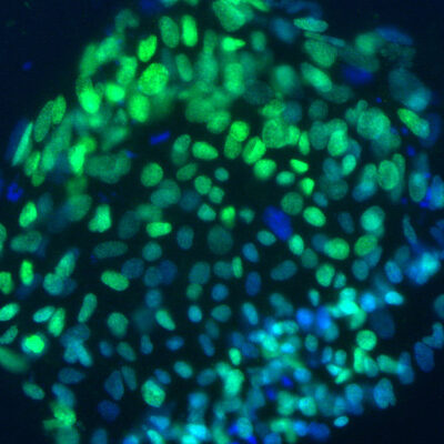
The University of Connecticut Cell and Genome Engineering Core has substantially contributed to Stem Cell Research conducted in Connecticut and beyond by providing a central source of technologies and materials for research on human pluripotent stem cells: embryonic stem cells (hESC) and induced pluripotent stem cells (hiPSC). The Genome Editing arm of the Core uses cutting edge genome engineering technologies to develop cellular models.
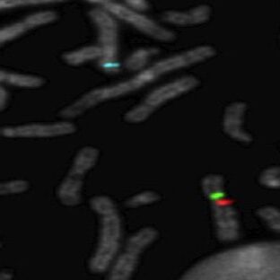
The mission of the CGI is to provide state-of-the-art expertise in genome technologies while facilitating genomics research for faculty and students across the University of Connecticut campuses.
CGI's infrastructure serves as a nexus for computational biology support and maintains a diverse portfolio of genomics instrumentation from microfluidics, arrays and next generation sequencing. Support for these technologies are in the form of experienced user access, hands-on assistance, training and/or consultation through the CGI.

The Center for Mouse Genome Modification (CMGM) at UConn Health provides design and generation of genetically modified mice and other services.
Computational Biology Core (CBC)
Director
Jill Wegrzyn, Ph.D.
Associate Director (UCHC)
Vijender Singh, Ph.D.
The Computational Biology Core (CBC) housed in the Institute for Systems Genomics at the University of Connecticut provides computational power and technical support to UConn researchers and affiliates. We collaborate with the Center for Genome Innovation (CGI) and Proteomics & Metabolomics Facility (PMF, Storrs campus COR²E Facility) to provide full support to the research community.
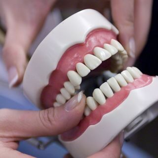
The Dental Clinical Research Center (DCRC) is a state-of-the-art center that supports oral health clinical research at UConn Health. The mission is to facilitate clinical research efforts of all faculty, as well as to encourage research collaborations among biomedical fields at UConn Health. Investigators are encouraged to contact the staff to discuss specialized needs.
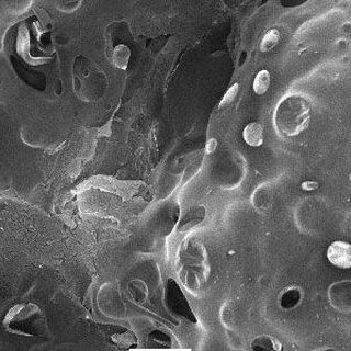
The Central Electron Microscope Facility (CEMF) is a UConn Health-supported research facility providing electron microscopy research service for faculty, students and extramural users. The CEMF occupies approximately 1,800 sq. ft. of space on B level of the main UConn Health building (rooms AB027 and AB031). The CEMF has three recently installed electron microscopes (EMs) equipped with digital imaging capabilities: a Hitachi H-7650 transmission EM, a JEOL JSM-5900LV scanning EM and a Zeiss Sigma Gemini HV field emission Scanning EM. Sample preparation instruments include ultramicrotomes, vacuum evaporator, sputter coater, critical point dryer, high pressure freezer, freeze substitution system and ion beam coater. The electron microscopes and other instruments also are available for use by experienced faculty or students. During the Fall of 2023, the facility will be installing a Thermo Fisher Tundra cryo-EM and a Zeiss Sigma 360 scanning EM equipped with an energy dispersive spectroscopy probe. These instruments will greatly enhance the capabilities of the EM facility.

The UConn Health Flow Cytometry facility provides flow cytometric analysis and cell sorting services to all UConn researchers as well as researchers at neighboring institutions. The UConn Health Center Flow Cytometry Facility has six instruments available for cellular analysis and two cell sorters. There are three Becton Dickinson (BD) LSR II instruments, an automated high-throughput Bio Rad ZE5, an Accuri C6 analyzer, and an Amnis ImageStream Mark II. Cell sorters include two BD FACS ARIA II high speed cell sorters.
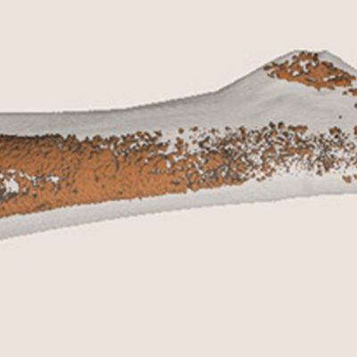
The microCT Imaging Facility specializes in quantitative 3-dimensional imaging using x-rays. Our most common projects involve morphometric and densitometric analyses of mineralized tissues from model organisms. In addition to bones and teeth, we provide nondestructive scans of biomaterials, museum artifacts, and industrial products with micrometer-scale resolution. We operate 3 Scanco µCT instruments and have an arrangement of micromechanical testing devices to allow detailed assessment of structure and function.
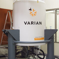
The NMR Facility provides a state-of-the-art environment for studying the structure, dynamics, folding, and interactions of biological macromolecules. The primary goal of the facility is the application of advanced NMR techniques to important and challenging biomedical problems. To meet these challenges, facility staff adopt state-of-the-art methods and develop new methods to enhance and facilitate bio-molecular applications of NMR, including structural biology, interaction screening, and metabolomics.
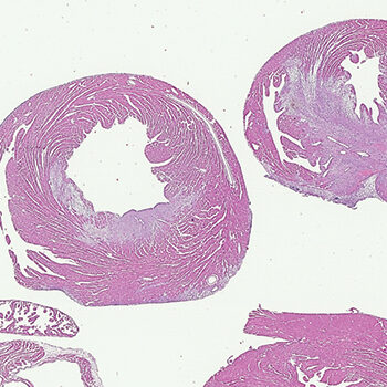
The UConn Health Research Histology Core is a fee-for-service facility dedicated to providing histology support for research investigators.
Proposal Details
- The program is for new projects that expand the capabilities of a core or lead to new preliminary data for a new grant. Projects already funded on an existing grant are not eligible.
- Award amounts vary by core and are capped at $8,000.
- Awards can only be used for core service charges. They cannot be used to support personnel or supplies in the PI's laboratory.
- Projects should be completed within 6 months of funding.
Proposal Evaluations
- Core directors will select the proposals they wish to support and will submit an application to HCRAC on your behalf by Friday, September 1, 2023.
- HCRAC will select finalists and may invite them to give a brief presentation on Thursday, September 21st.
- Final decisions will be made in early October.